Dr Chin’s Guide to Discover Ear Anatomy: Key Parts for Balance and Sound
Discover the essentials of ear anatomy and learn how its key parts contribute to balance, sound, and speech in our informative blog post.
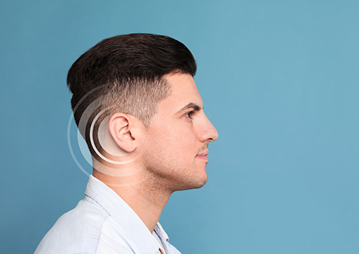
The ear is a complex system located on the side of the head, consisting of the external ear, tympanic cavity (middle ear), and internal ear. Its functions include sound perception, balance, and speech development. Sound waves enter the ear through the external auditory meatus, which travels the ear canal, triggering vibrations that stimulate the cochlear duct. The auditory tube maintains pressure balance, while the tensor tympani muscle dampens loud sounds. The internal acoustic meatus carries the facial nerve and vestibulocochlear nerve, which detects motion and linear acceleration. Blood flow to the ear comes from various branches, and sensory innervation is provided by Arnold’s nerve.
The Outer Ear: Directing Sound Waves Inward
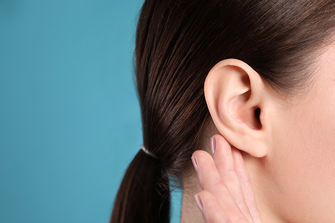
The outer ear is the visible part of the ear and the entryway for sound.
- Pinna (Auricle): This external structure collects sound waves from the environment and funnels them into the auditory canal. Its unique shape enhances directional hearing.
- External Auditory Canal: The canal carries sound waves toward the eardrum. It also produces cerumen (earwax), which traps debris and protects the inner structures.
This portion of the ear is essential for localization of sound and basic sound amplification.
The Middle Ear: Amplifying Vibrations
The middle ear lies behind the eardrum and is filled with air. It contains three bones collectively called the ossicles—the malleus (hammer), incus (anvil), and stapes (stirrup).
- Tympanic Membrane (Eardrum): Vibrates in response to sound waves.
- Ossicles: Transmit and amplify those vibrations to the oval window of the inner ear.
- Eustachian Tube: Connects the middle ear to the back of the nose and throat, equalizing pressure and preventing fluid build-up.
The Inner Ear: Processing Sound and Controlling Balance
The inner ear is responsible for converting sound into nerve signals and maintaining balance. It houses two major components: the cochlea and the vestibular system, both encased in the bony labyrinth of the skull.
Cochlea: Hearing’s Command Center
The cochlea is a spiral-shaped, fluid-filled organ responsible for converting sound waves into electrical signals.
- Inside, thousands of tiny hair cells detect different frequencies.
- These signals are sent via the auditory nerve to the brain for interpretation.
Damage to hair cells can lead to sensorineural hearing loss, which is often permanent.
Vestibular System: The Balance Regulator
Made up of the semicircular canals, utricle, and saccule, this system detects motion, head position, and gravitational pull.
- Semicircular Canals: Detect rotational movements.
- Utricle and Saccule: Detect vertical and horizontal movements.
Disorders in this system can lead to vertigo, imbalance, and motion sensitivity.
Why ENT Evaluation Matters
When patients present with ongoing ear pain, hearing loss, severe vertigo, or speech delays, ENT evaluation is critical. Conditions like otitis media, swimmer’s ear, or tympanic membrane perforation may involve structures like the ear drum, cochlear nerve, or vestibular nerve, housed in the temporal bone. Early diagnosis prevents complications such as permanent hearing loss, endolymphatic hydrops, or benign paroxysmal positional vertigo—all well-documented in StatPearls Publishing and otolaryngol head neck surg literature.
- About the Author
- Latest Posts

Dr Ronald Chin is an Australian trained Otolaryngologist Head and Neck Surgeon.
After graduating as a Fellow of the Royal Australasian College of Surgeons, Dr Chin undertook further specialised training in Head and Neck Cancer at the Royal College of Surgeons in Ireland.
He has published many research papers and is an active teacher and scholar.
As part of his subspecialty training, Dr Chin has training in Laser, Da Vinci Robotic, Flex Robotic and complex surgical techniques.
In addition to specialised Head and Neck Cancer, Dr Chin also enjoys general adult and paediatric ENT Surgery and practices sinus, snoring/sleep and general paediatric ENT Surgical procedures.
Dr Ronald Chin works as a general Otolaryngologist, offering a wide range of surgical and non-surgical treatments including ear surgery, nose surgery and throat surgery. He provides treatment for chronic conditions such as tonsillitis, sinus problems and problems with hearing.
He is also involved in the diagnosis and treatment of many conditions such as facial paralysis, head and neck cancer and sleep apnea. As well as performing surgery on children, he also provides specialist care for adults, including the treatment of throat disorders, voice loss and ear problems.
Dr Chin has also served as a Conjoint Associate Professor at the University of Sydney, a Conjoint Associate Professor at Western Sydney University and an Adjunct Associate Professor at the University of Technology Sydney.